Microscopy in the age of quantum physics
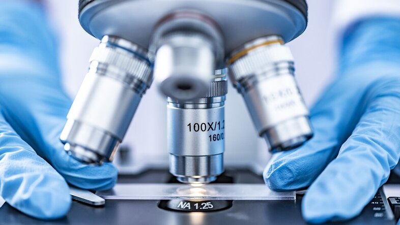
Much lies beyond what we can see with the naked eye: bacteria, viruses, atoms. Antonie van Leeuwenhoek was the first to see bacteria through a microscope 350 years ago – a "small" discovery that revolutionised biology and medicine. Today, thanks to quantum physics, we can see individual atoms. In biology, microscopy now faces the challenge of imaging molecular dynamics.
Using traditional light microscopy, we can visualise structures that are larger than the wavelength of light. For a long time, it seemed impossible to see, let alone manipulate, individual atoms. Quantum physics laid the foundations of new microscopy methods, which are now once again enabling major advances in science and technology.
Quantised energy levels
One of these foundations is the discrete energy levels of atoms and molecules, as described by quantum physics. These are essential for fluorescence microscopy, which enables us to detect individual molecules. By skilfully exploiting the energy levels and their temporal dynamics, it is possible to surpass the limit for microscope resolution. Super-resolution microscopy has led to countless discoveries in cell and molecular biology.
Using electrons to see atoms
Another foundation of modern microscopy is that electrons are described by a wave function in quantum physics. Similar to electromagnetic waves (light), these so-called matter waves can be diffracted off objects or focussed with a lens. However, their wavelength is in the picometre range, that is a millionth of a millionth of a metre. This motivated Ernst Ruska to build the first electron microscopes in the 1930s. He soon achieved a resolution that was 100 times better than that of light microscopy. Electron microscopes today achieve a resolution of a few tens of picometres. This allows us not only to see individual atoms but also to manipulate them, which makes electron microscopy indispensable in nanophysics and materials science.
Good resolution alone is not enough
However, this resolution has not yet been achieved in biological applications. Since biological samples are destroyed by interaction with electrons, only a few electrons can be used for imaging. This results in noisy images. A major breakthrough was achieved with the development of cryogenic electron microscopy, in which tens of thousands of noisy images of identical proteins are analysed to find the atomic structure of this protein. Among other things, this method helped researchers to decipher the structure and function of different variants of the COVID-19 virus.
However, if we want to image a single protein in high resolution, or image the dynamics of proteins, it is essential to develop new microscopy methods with higher sensitivity. Quantum research can contribute significantly to this. In the following, I would like to describe three approaches from my lab.
Controlling electrons with light
One approach is to optimise the electron microscope for the investigation of a specific sample. In my lab, for example, we make use of the interaction between electrons and light to manipulate the wave function of the electrons. The aim is to shape the wave function in such a way that the information obtained via electron-sample interaction is maximized.
Reading light using electrons
In another project, we are making use of the photoelectric effect to combine the benefits of light microscopy and electron microscopy. In cooperation with Leiden University and the Heyrovsky Institute, we have built the world's first optical near-field electron microscope: We use light to illuminate a sample and convert the resulting light intensity patterns into an electron beam, which we image. This is done very close to the sample, which allows us to achieve a high spatial (~30nm) and temporal (<0.1s) resolution. The sample remains intact and does not need to be chemically modified. This offers unique opportunities for investigating dynamic processes on surfaces – whether in electrochemistry or biophysics.
Entangled electrons
Finally, I would like to talk about entanglement. This special type of correlation between two particles can be used to make measurements more precise. This is already being done to improve sensitivity in gravitational wave detection. Together with colleagues from the TU Wien, the JKU in Linz and the University of Innsbruck, we try to entangle electrons with the ions of a quantum computer. In the first step, this will help us gain a better understanding of the interaction between an electron and an atom. In the second step, we want to use entanglement to gain more information about a sample before it is destroyed. In the future, this could help to reconstruct the structure of individual proteins or to better visualise the architecture of cells. This would be a further step towards microscopy methods that make biology visible at the molecular level.
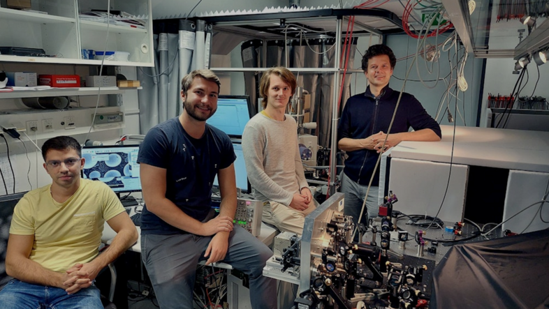
The group aims to develop new methods that can image dynamic samples with high resolution and sensitivity. They work to translate concepts from quantum metrology to microscopy. The microscopes developed are applied in a wide spectrum of fields, ranging from biophysics to electrochemistry.

